Bacteria
Bacteria are single-celled microorganisms that impact your daily life. Your body has many different kinds of bacteria that perform vital metabolic functions and provide key protection against foreign substances. But, bacteria can also be pathogenic (disease-producing).
Of all living groups, bacteria alone are prokaryotic. Prokaryotic organisms do not have membrane-covered nuclei or cellular organelles. This makes them very unique among the many species on Earth, and being prokaryotic does not mean they are that “simple”. They have a remarkable reproductive capability and metabolic diversity. They survive in deserts, hot springs, glaciers, and seas and in the organisms that live in those environments.
Bacteria were the first living organisms and continue to exist in close relationship to many other species.
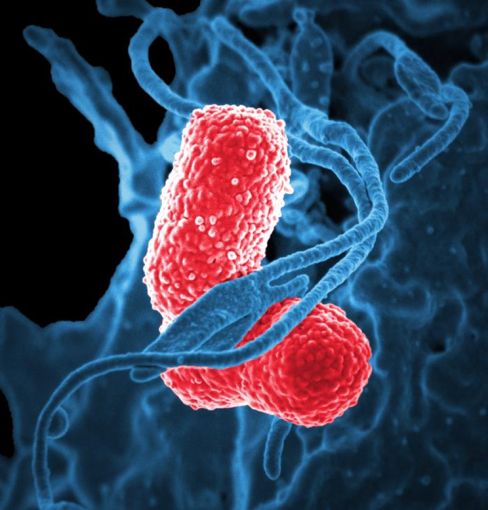
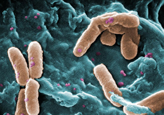
Image: This digitally enhanced image of a scanning electron microscope view shows a human white blood cell with two rod-shaped Klebsiella pneumonia bacteria known to cause severe infections.
Photo Credit and Source: David Dorward; Ph.D.; National Institute of Allergy and Infectious Diseases (NIAID)
Objectives
To understand the importance of bacteria and bacterial characteristics, you should be able to:
- Know how bacteria are classified.
- Identify key bacterial structures and their functions.
- Explain different mechanisms by which bacteria get energy.
- Distinguish between prokaryotic and eukaryotic cells.
- Describe how bacteria are classified by shape.
- Explain how bacteria can be identified by using a stain.
Vocabulary
- bacteria–one-celled, prokaryotic organisms which are involved in fermentation, nitrogen fixation, and infectious disease.
- bacillus–rod-shaped bacteria.
- coccus–round-shaped bacteria.
- eukaryotic–an organism whose cells have specialized, membrane-covered organelles.
- fission–a form of asexual reproduction; the splitting of an organism into two parts.
- flagellum–a structure that allows cells to be motile; usually has a core of 9 + 2 array of microtubules.
- Gram stain–a type of stain used to identify bacteria as being either Gram positive or Gram negative, depending on the chemical characteristics of the bacterial cell wall.
- microorganism–an organism too small to be seen without a microscope.
- prokaryotic–a single-celled organism which lacks membrane-covered organelles.
- saprobe–heterotrophs that obtain energy and carbon from non-living organic matter, causing decay).
- spirillum–spiral-shaped bacteria belonging to the genus of Gram-negative bacteria.
Getting Energy
All organisms must take in energy to survive and perform metabolic and reproductive activities. Bacteria differ in the way they obtain energy.
photoautotrophs
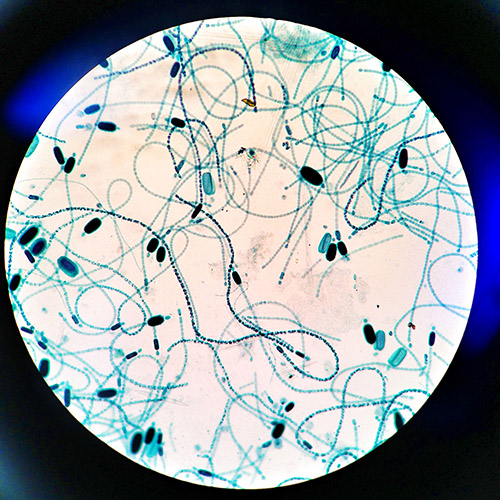
Cylindrospermum, a cyanobacteria capable of photosynthesis.
Image source: Wikipedia
Photoautotrophs are bacteria that are photosynthetic. They use sunlight for energy and carbon dioxide as a carbon source.
chemoautotrophs
Venenivibrio stagnispumantis is a chemoautotroph which obtains energy by oxidizing hydrogen gas.
Image source: Wikipedia
Chemoautotrophs synthesize their own organic compounds using only carbon dioxide as the carbon source and an inorganic substance as an energy source; they obtain energy from chemical reactions.
photoheterotrophs
Bacteria that use the sun’s energy for photosynthesis and fatty acids, complex carbohydrates, and other compounds as carbon sources are photoheterotrophs. Unlike photoautotrophs, they cannot use carbon dioxide as their sole carbon source.
chemoheterotrophs
Chemoheterotrophs are bacteria that are parasites or saprobes. Parasites live on or in a living host and saprobes get nutrients from wastes or remains of other organisms. They cannot use carbon dioxide to form their own organic compounds.
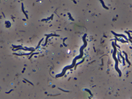
Bacterial Shapes
Bacteria have three basic shapes: spherical, rod-shaped, and spiral-shaped. A bacterium with a spherical shape is a coccus (plural, cocci), one having a rod shape is a bacillus (plural, bacilli), and one with twists is a spirillum (plural, spirilla).
There are a variety of shapes within the three groups. Cocci can be somewhat flattened, bacilli can be tapered at the ends, and spirilla can be comma-shaped or have multiple twists.
Bacteria are unicellular and prokaryotic, meaning that they do not have membrane-covered organelles which separate chemical reactions inside the cell. Metabolic reactions take place in the cytoplasm.
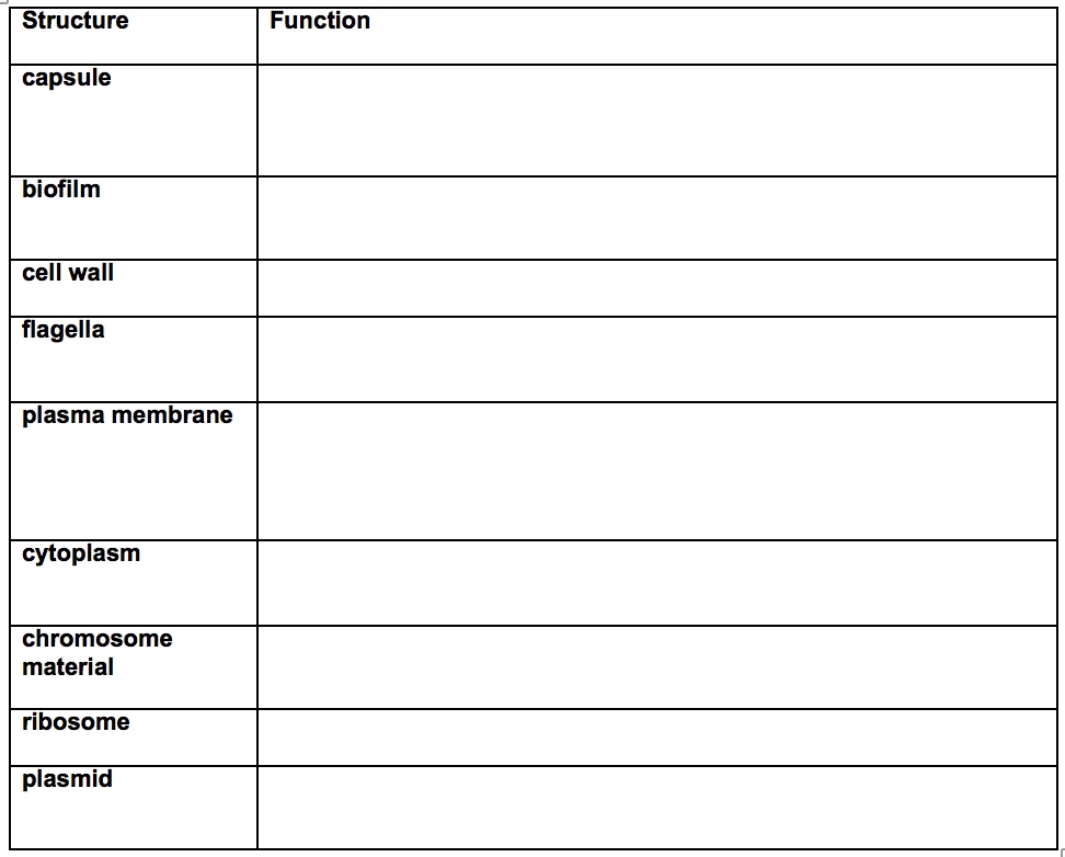
Bacterial Structures
1) capsule–a sticky layer outside the cell wall that allows some bacteria to stick to surfaces such as teeth and mucous membranes. It also helps some bacterial species avoid being engulfed by the body’s infection-fighting cells and helps prevent the bacteria from drying out in some environments.
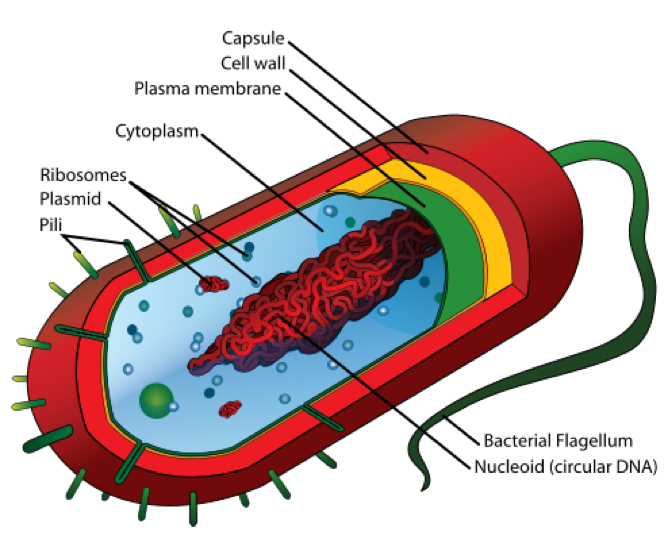
Image source: Wikipedia
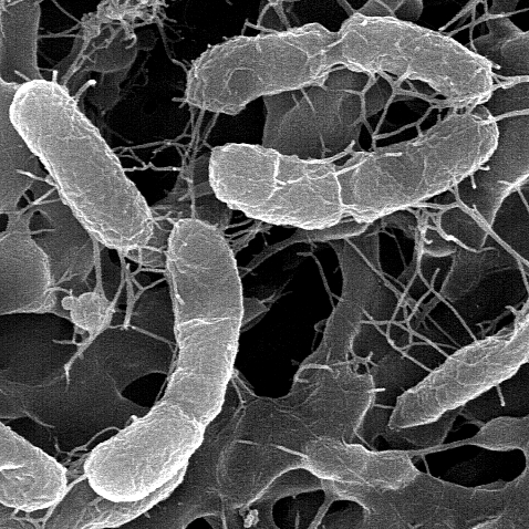
The capsule can also have a slime layer or biofilm. The slime layer allows the bacteria to stick to materials and each other to form microcolonies within the slime layer. Bacterial biofilms are important considerations in some health conditions. Streptococcus mutans grows a biofilm that can decalcify tooth enamel and leads to infections of the teeth. Pseudomonas aeruginosa biofilms cause chronic lung infections in patients with cystic fibrosis and make the infections much harder to treat.
This image shows a scanning electron micrograph image of Pseudomonas aeriginosa.
Image source: Wikipedia
Credit: Janice Haney Carr Permission: PD-USGov-HHS-CDC
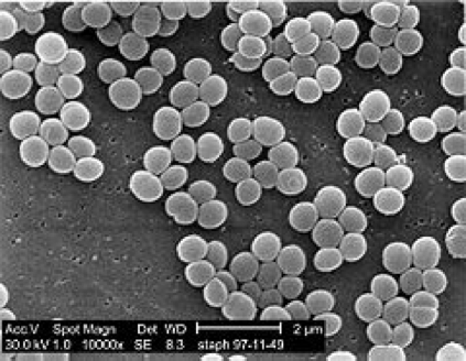
For example, to the right is an image of Staphylococcus aureus on the surface of an indwelling catheter. The sticky-looking substance between the round-shaped bacteria (cocci) is the bacterial biofilm. The biofilm protects the bacteria and makes the infection more difficult to treat with antibiotics.
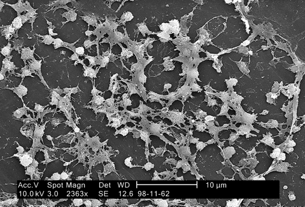
Image: Wikipedia
Photo Credit: Janice Carr Permission: PD-USGov-HHS-CDC
2) cell wall–a semi-rigid permeable structure just inside the capsule that surrounds other bacterial structures and allows the bacteria to keep its shape. It is made up of peptide (protein) groups and polysaccharide (sugars) strands.
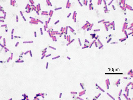
The characteristic peptides and polysaccharides allow scientists to identify a major characteristic of bacteria using a purple stain called Gram’s stain. Depending on the chemical compounds, bacterial cell walls either take up the purple stain after an iodine wash (called Gram-positive) or they do not take up the purple stain (called Gram-negative). This characteristic of the cell wall is a major tool used by clinicians to identify the bacteria in a bacterial culture.
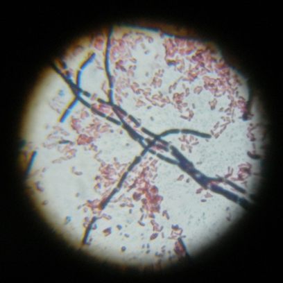
The image at the left as seen through a microscope shows both Gram-positive (stained dark purple) rod-shaped bacteria, Bacillus cereus, and the smaller Gram-negative rod-shaped bacteria E. coli (stained pink).
A few bacteria do not have cell walls. They have, instead, chemical compounds which protect the bacteria against drying out and they usually live in environments in which osmotic pressures are not a problem for survival. Mycoplasma pneumoniae is an example of a cell wall-less bacteria. Because it has no cell wall it can have a variety of shapes.
3) flagella–a whip-like structure used for movement. Some, but not all, bacteria have one or more flagella. These structures move in a circular motion to propel the bacteria forward. Bacteria can have multiple flagella that surround the cell, a few flagella on one or both ends of the cell, or a single flagella.
Using flagella, bacteria are able to respond to environmental stimuli. They are able to sense the presence of certain nutrients in their environment and move toward them and they are able to move away from harmful substances.
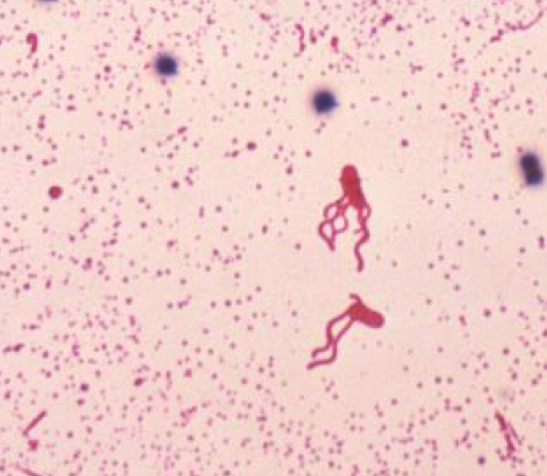
Two flagellated Bacillus coagulans bacteria stained with Leifson flagella stain method appear in this blood smear.
Image Credit: CDC/Dr. William A. Clark.
Of medical significance, some bacteria use their flagella to propel themselves into body organs through tubes and ducts. Urinary tract infections can become bladder infections after bacterial flagella propel the bacteria up the urethra into the bladder. Some organisms can be identified by their motility (movement) pattern in cultures.
4) plasma membrane–a membrane surrounding the cytoplasm just inside the cell wall. The plasma membrane provides support and allows movement into and out of the cell. Its main function is providing the selective permeability that allows metabolic function. It also participates in energy-providing functions, synthesis of membrane lipids, and motility functions.
5) cytoplasm–a gel-like substance inside the plasma membrane, it is the site of metabolic and reproductive processes.
6) ribosomes–site for protein synthesis. These structures are made up of RNA and protein.
7) chromosome material–genetic material of the cell. Bacterial chromosome material is DNA.
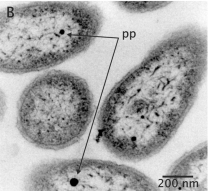
8) plasmids–extrachromosomal genetic material; made up of DNA.
Check Your Understanding
- Name and describe three bacterial shapes.
- Describe 4 ways bacteria get energy.
- What is a bacterial biofilm and what is its significance in infectious disease?
- Complete the following table:
- Label the following diagram of bacterial structure.